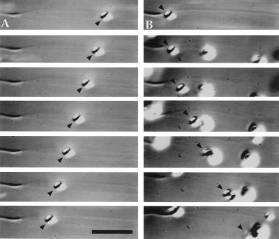
Figure 2.
Bidirectional motility of a single melanosome along an axoneme-nucleated microtubule observed by video-enhanced DIC microscopy. Portions of a video sequence showing (A) minus end-directed motility and (B) plus end-directed motility of the same melanosome (arrowheads). The elapsed time between frames is 2 s. (Bar = 6 μm.)

