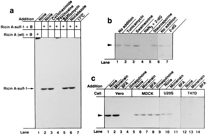
Figure 2.
Sulfation of ricin A-sulf-1 upon incubation of reconstituted toxin with different cells. (a) Near confluent Vero cells growing in 5-cm2 dishes were washed twice with DMEM without sulfate and incubated with the same medium containing 100 μCi/ml Na235SO4 for 3 h. The indicated compounds were added, and after 30 min, 200 ng/ml reconstituted ricin containing ricin A-wt (lane 1) or ricin A-sulf-1 (lanes 2–7) was added and the incubation was continued for 4 h. Then the cells were washed with PBS and lysed, the nuclei were removed by centrifugation, and the clear supernatant was submitted to immunoprecipitation with rabbit anti-ricin antibodies immobilized on CNBr-Sepharose 4B. The adsorbed material was analyzed by SDS/PAGE under reducing conditions. The additions were as follows: lane 3, 20 μg/ml cycloheximide; lane 4, 20 μM psychosine; lane 5, 0.1 μM bafilomycin A1; lane 6, 30 μM nocodazole; lane 7, the incubation temperature was 17°C. (b) Conditions were as in a, lane 2, but the additions were as follows: lane 2, 1 μM tunicamycin; lane 3, 1 μg/ml swainsonine; lane 4, 10 mM NaN3 and 50 mM 2-deoxyglucose; lane 5, 0.1 μM staurosporil; lane 7, 50 mM 2-deoxyglucose. (c) Cells as indicated were treated as in b with the following additions: lanes 2, 6, 10, and 13, 10 μM monensin; lanes 3, 7, 11, and 14, 2 μg/ml brefeldin A; lanes 4 and 8, 30 μM ilimaquinone.

