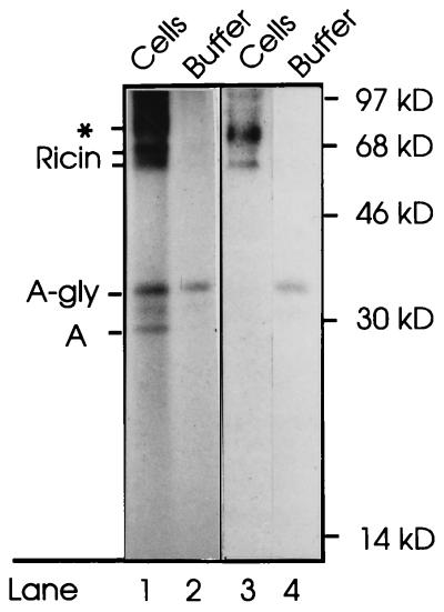
Figure 6.
Translocation of ricin A chain to the cytosol. Vero cells were incubated with Na235SO4 and ricin containing A-sulf-2 for 4 h. Then N-ethylmaleimide (10 mM) was added and the cells were further incubated for 10 min and then washed twice with PBS containing 0.1 M lactose. The cells were permeabilized with SLO. After collection of the buffer, the cells were lysed in Triton X-100 and centrifuged to remove the nuclei. Both supernatant fractions were treated with immobilized anti-ricin, and the adsorbed material was submitted to SDS/PAGE under nonreducing conditions. Lanes 1 and 2, and lanes 3 and 4 represent two different experiments. On the left, the positions of glycosylated and unglycosylated ricin holotoxin, and of glycosylated and unglycosylated free ricin A-sulf-2 are indicated. The band marked by an asterisk indicates the position of a contaminant, presumably a sticky sulfate-containing proteoglycan, that often comes down together with the immunoprecipitate. Molecular mass markers are indicated on the right.

