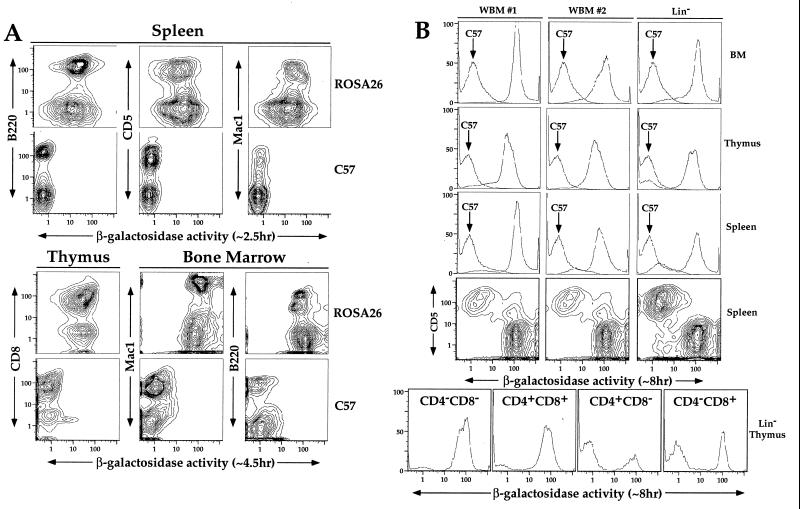
Figure 2.
Multiparameter FACS-Gal analysis of hematolymphoid organs in ROSA26 mice and mice transplanted with ROSA26 bone marrow. (A) Multiparameter FACS-Gal analysis of adult hematolymphoid organs in ROSA26 homozygous mice. Nucleated cell suspensions from spleen, thymus, and BM of ROSA26 homozygous mice were loaded with FDG, stained with antibody markers specific for certain hematopoietic lineages (B cells, B220; T cells, CD5 or CD8; myeloid cells, Mac1) and analyzed by FACS. To determine background fluorescence in the FACS-Gal assay, fluorescence levels of ROSA26 mice were compared with C57BL/6J controls (contour plots labeled C57 on right). (B) FACS-Gal analysis of C57BL/6J mice transplanted with ROSA26 BM cells. After lethal irradiation, mice were transplanted with either whole BM (WBM #1 or #2) or cells sorted to be negative for a panel of hematolymphoid lineage markers (Lin−). Four weeks after irradiation and transplantation, single cell suspensions were prepared from BM, thymus, and spleen of the transplant recipients; loaded with FDG; stained with various antibody markers; and analyzed on the FACS. To assess the degree of reconstitution by the ROSA26 bone marrow cells, the fluorescence histogram of an identically prepared cell suspension from C57BL/6J age-matched mice is denoted by an arrow (C57). To demonstrate that the remaining host cells found in the spleen are T cells (high CD5 expressing cells), we show two-color probability plots for CD5 vs. β-gal staining of splenocytes. To determine which stages of thymocyte development still contain a significant number of host cells, we show FACS-Gal histograms electronically gated for various patterns of CD4 and CD8 expression in the thymus of the mouse reconstituted with Lin− cells (Lin− Thymus).

