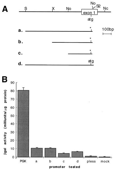
Figure 6.
Characterization of the ROSA26 promoter. (A) Map of the genomic DNA at the 5′ end of exon 1 of transcripts 1 and 2. The relative position of fragments used for promoter activity assay is shown. (a–c) A potential translation start site in exon 1 (atg) has been converted to a BamHI site (asterisk). Nc, NcoI; No, NotI; S, SalI; S2, SacII; X, XhoI. (B) The various promoter fragments (bars a–d) were fused to lacZ, the constructs were electroporated into ES cells, and β-gal activity was assayed. The PGK promoter (PGK) was used as a positive control and no fragment (pless) and mock-transfected cells (mock) were used as negative controls.

