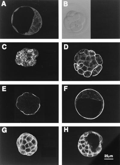
Figure 3.
Cellular localization of GLUT3 and GLUT1 during mouse preimplantation development. Confocal images of optical sections of early four-cell, six-cell embryos, early morula, compacted morula, and blastocyst incubated with 10 μg/ml anti-GLUT3 antibody (B–F); morula and blastocyst incubated with anti-GLUT1 antibody (G and H); and blastocyst incubated with pooled nonimmune rabbit serum (A). GLUT3 protein is first apparent at the late four- to six-cell stage (C) when it appears restricted to cytoplasmic vesicles. Note that, as the embryo develops, positive immunoreactivity first appears on plasma membranes then concentrates in the apical surface of the polarizing outer cells of the compacted morula. In blastocysts it is restricted to the apical membranes of TE. This contrasts with GLUT1 expression which is localized in basolateral membranes of both morulae (G) and blastocysts (H).

