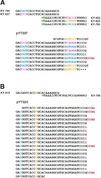
Figure 3.
MPR with mixtures of primers. (A) Five primers used in MPR are shown on the top. Mismatched nucleotides are in green, and sequences that are differ among primers are separately colored. MPR was carried out with an Expand Taq polymerase mixture for 45 cycles at 69°C for extension. An example of a microgene polymer (pYT037) is shown on the bottom. Inserted nucleotides or deletion (Δ) at junctions is in red. (B) A degenerated primer was used in MPR. Ns represent a mixture of A, T, G, and C. MPR was carried out with an Expand Taq polymerase mixture for 65 cycles at 69°C for extension. An example of a polymers (pYT020) is shown.

