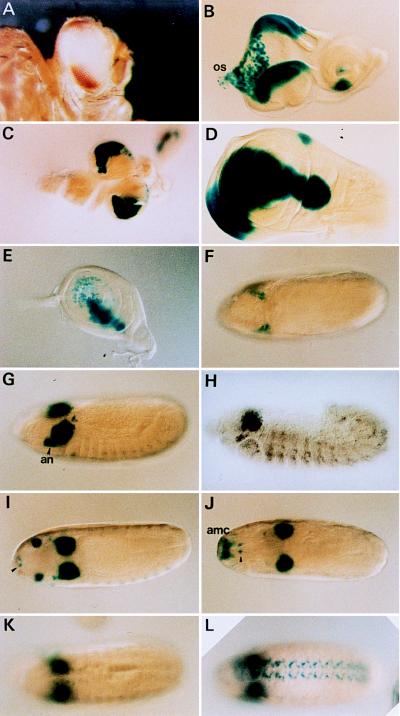
Figure 1.
Expression patterns of reporter genes in ombP1 (A–G and I–K) ombP1, (H) wild type, (L) D13–1. (A) The bipolar w+m expression pattern in ombP1. (B–G and H–K) lacZ expression pattern as stained by X-Gal. The pattern is similar to omb expression (3, 29), except when noted below. (B) In eye–antennal disc expression is in the dorsal and ventral poles of the eye disc, primarily in the peripodial membrane destined to become part of the head capsule (30). Expression is also seen in scattered cells in the proximal region of the disc and the optic stalk (os), and in a ventral sector in the antennal disc. (C) Expression in the central nervous system is in the optic lobe, plus a few cells in the ventral ganglia. (D) Expression in the wing disc is in a broad domain straddling the anterior–posterior compartmental boundary. This expression has recently been shown to be under dpp and wg regulation (29, 31, 32). Weak omb expression in the notal region is not detected in lacZ expression (compare with figure 2C in ref. 29). (E) In the leg disc, expression is also in a stripe along the anterior–posterior compartmental boundary, but only in the dorsal region. (F) Dorsal view of a stage 8 embryo. Expression is in two lateral clusters of cells anterior to the cephalic furrow. (G) Slightly oblique lateral view of a stage 12 embryo. Expression is strong in the optic lobe anlage and antennal segment (an). The lateral segmentally repeating pattern is weaker than omb (compare with H). (H) Expression detected by in situ hybridization of omb in a wild-type embryo of similar stage as in G. The antennal expression is weaker. (I) Dorsal view at about stage 13 (before head involution); the antennal expression had moved dorsally and anteriorly. Expression also occurred in two ventral anterior spots, probably the hypopharyngeal sensory organ (ho; arrowhead). (J) After head involution, the antennal expression spot had moved to a position corresponding to the antennomaxillary complex (amc) (dorsal view). (K) X-Gal staining in the ventral nerve cord is weaker compared with omb in situ hybridization in wild type (compare with figure 2H in ref. 3). (L) The ventral nerve cord expression is stronger in D13–1. (K and L) Dorsal view.

