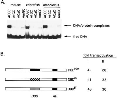
Figure 4.
Recognition of Whn binding sites in vitro and in vivo. (A) Electrophoretic mobility shift assays using mouse, zebrafish, and amphioxus DNA binding domain peptides. A double-stranded oligonucleotide obtained via in vitro selection was used in wild-type configuration (core sequence ACGC), in modified forms (changed nucleotides in core sequence are indicated by lowercase letters), or in in vitro methylated form (m denotes 5-methylcytosine). (B) Transactivation of a luciferase reporter gene after transient transfection into BHK cells. A luciferase gene with a minimal promotor (11) was cotransfected with a mouse whn (DBDMm) expression plasmid or with constructs in which the mouse DNA binding domain was changed to that of zebrafish (DBDDr) or amphioxus (DBDBl) to establish a luciferase baseline activity. These values were compared with reporter constructs in which a whn response element was positioned upstream of the minimal promotor and expressed as fold transactivation. Values shown represent the average of two experiments with a variation of less than 20%. I refers to expression constructs with an N-terminal MYC tag; II refers to constructs with a C-terminal MYC tag. DBD, DNA binding domain; AD, transcriptional activation domain (8). In control experiments, the transfection of whn expression constructs in anti-sense orientation had no effect on luciferase activity.

