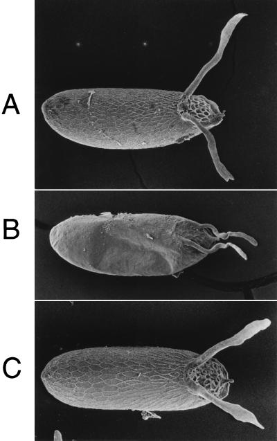
Figure 4.
Phenotypic rescue of the chorion defects in k43fs293. Laid eggs were analyzed by scanning electron microscopy for chorion morphology and are presented in a dorsal view, with anterior to the right. The two large chorion dorsal appendages are apparent at the dorsal anterior (right). (A) Wild-type Oregon R strain. (B) Mutant y ac w; k43fs293. (C) Mutant + rescue y ac w; p[yellow+; HX7.5]/+; k43fs293. The honeycomb-like pattern on the egg surface is the outline of chorion material deposited by each individual follicle cell. Note that in the mutant (B) the amount of chorion material present in the dorsal appendages and surrounding the egg is greatly reduced, and the mutant egg is slightly collapsed. All of these defects are corrected in the rescue strain (C).

