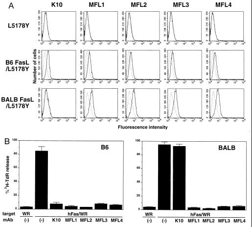
Figure 1.
Reactivity of anti-mouse FasL mAbs. (A) Cell surface staining of mouse FasL transfectants. L5178Y, B6 FasL/L5178Y, and BALB FasL/L5178Y cells were stained with anti-mouse FasL mAbs, K10, and MFL1∼4, followed by FITC-labeled goat anti-mouse IgG antibody that crossreacts with hamster IgG (solid lines). Broken lines indicate background staining with FITC-labeled goat anti-mouse IgG antibody alone. (B) Blocking of cytotoxic activity of mouse FasL transfectants. Cytotoxic activities of B6 FasL/L5178Y or BALB FasL/L5178Y cells were tested against hFas/WR19L (hFas/WR) or WR19L (WR) target cells in the absence or presence of 10 μg/ml anti-mouse FasL mAbs (K10 and MFL1∼4) in a 6-h 3H-TdR release assay at an E/T = 10. Data represent mean ± SD of triplicate samples.

