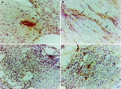
Figure 3.
In situ PCR detection of parasite kDNA in native and transplanted hearts. Detection of a parasite-infected cell (pseudocyst) in the heart of a mouse 14 days postinfection (A) demonstrates the specificity of the in situ PCR for the parasite. Native heart tissue from chronically infected mice (350 days) shows no intact parasite-infected cells but displays a diffuse staining pattern that is more intense in areas of heavy inflammation (arrows in B). The transplanted heart present for 200 days in the same animal as in B shows no evidence of either intact or diffusely distributed parasite kDNA, nor any evidence of inflammation (C). Five days after injection of 106 parasites into a transplanted heart, parasite kDNA is readily detected by in situ PCR (D). The detection of kDNA in the parasite-injected transplanted heart precedes the inflammatory response evident by day 15 postinjection (see Fig. 2). (×250.)

