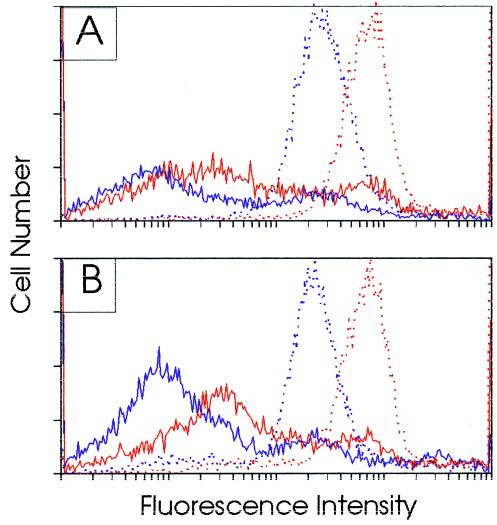
Figure 4.
Detection of fluorescein-labeled thioate and amidate c-myc AS-ODNs in normal splenocytes (CD45− cells) and in HL-60 cells (CD45+ fraction) in vivo. Spleen (A) and bone marrow (B) cell suspensions obtained 12 hr after the last injection of a 10:1 mix of unlabeled and fluorescein-labeled ODNs were incubated with a phycoerythrin-conjugated anti-CD45 monoclonal antibody. The fluorescence of the CD45+ and the CD45− fractions were determined by fluorescence-activated cell sorting analysis. Solid red line indicates fluorescent phosphoramide ODN in CD45− cells; dotted red line indicates fluorescent phosphoramide ODN in CD45+ cells. Solid purple line indicates fluorescent phosphorothioate ODN in CD45− cells; dotted purple line indicates fluorescent phosphorothioate ODN in CD45+ cells.

