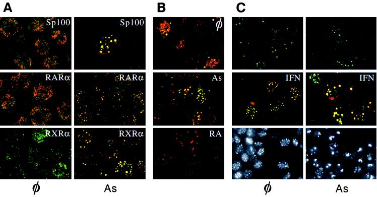
Figure 1.
(A) Confocal laser microscopy analysis of NB4 leukemic cells double-labeled with anti-PML antibodies [revealed by fluorescein isothiocyanate (FITC)] and anti-Sp100 (Top), anti-RARα (Middle), and anti-RXRα (Bottom) antibodies (revealed with Texas Red). Cells were treated (Right) or not (Left) with 10−6 M arsenic trioxide for 5 h. (B) Differential effects of RA and As2O3 (10−6 M, 24 h) on PML/RARα localization in CHO PML–PML/RARα cells. Double-staining with anti-PML (Texas Red) and anti-RARα (FITC) antibodies. (C Top and Middle) Arsenic recruits PML and Sp100 onto NB in HeLa cells. Double anti-PML (FITC)/anti-Sp100 (Texas Red) staining. Cells were primed (or not) with 500 units/ml IFNα for 12 h and treated (As) or untreated (⊘) with 10−6 M As2O3 for 7 h. (C Bottom) Immunofluorescence analysis of CHO PML cells. φ, untreated; As, arsenic-treated for 12 h. Note that arsenic induces the disappearance of the diffuse nuclear staining.

