Figure 5.
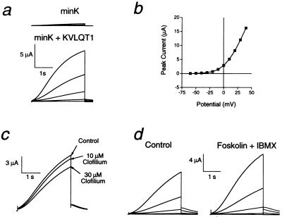
Functional and pharmacological characterization of minK+KvLQT1 currents in Xenopus oocytes. (a) Families of currents from minK- and minK+KvLQT1-injected oocytes elicited by 3-s voltage steps from a holding potential of −80 mV to test potentials ranging from −40 to +40 mV (20 mV increments). Voltage was returned to the original holding potential after the voltage steps. Currents were recorded 3 days after injecting cRNAs. Peak outward current amplitudes at +40 mV in minK-injected and minK+KvLQT1-injected oocytes were 0.5 and 10.3 μA, respectively. (b) Peak current voltage relationship for six oocytes expressing minK+KvLQT1. Currents were elicited 4 days after injecting oocytes by 3-s voltage steps from −80 mV to potentials ranging from −60 to +40 mV. (c) Effects of clofilium on minK+KvLQT1 current. Superimposed currents were recorded during 3-s steps to +30 mV from −80 mV during the same experiment. Clofilium was applied via bath perfusion. (d) Effects of forskolin and IBMX on minK+KvLQT1 currents. Currents were recorded during 3-s voltage steps from −80 mV to potentials between −50 and +30 mV (20 mV increments). Tail currents were elicited by stepping back to −70 mV. Currents were recorded before and 10 min after adding 10 μM forskolin and 100 μM IBMX to the bath.

