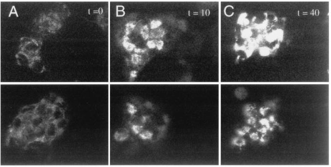
Figure 1.
Confocal photomicrographs of DHR fluorescence in GT1-1 trk cells in the absence (Upper) or presence (Lower) of NGF. Fluorescent images obtained immediately after loading (A, t = 0) show low basal fluorescence. Cells were then treated with vehicle (media; Upper) or NGF (100 ng/ml; Lower), and follow-up images were obtained at 10 min (B) or 40 min (C). A substantial increase in staining with rhodamine 123, the fluorescent oxidation product of DHR, can be seen in the absence of NGF but not in the presence of NGF.

