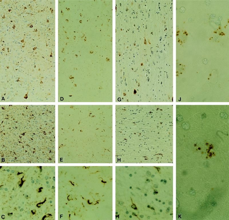
Figure 1.
Temporal cortex from a familial MSTD patient showing neuronal and glial cells stained with phosphorylation-dependent anti-tau antibodies. Neurons and glial cells in grey matter stained with antibodies AT8 (A), AT180 (D), and PHF1 (G). Antibody 12E8 stains granular deposits in neurons and glial cells in both grey (J) and white matter (K). In white matter AT8 (B and C), AT180 (E and F), and PHF1 (H and I) stain numerous glial cells with cytoplasmic inclusions (C, F, and I). (Magnifications: A, B, D, E, G, and H, ×140; C, F, and I, ×448; J, ×760; and K, ×112.)

