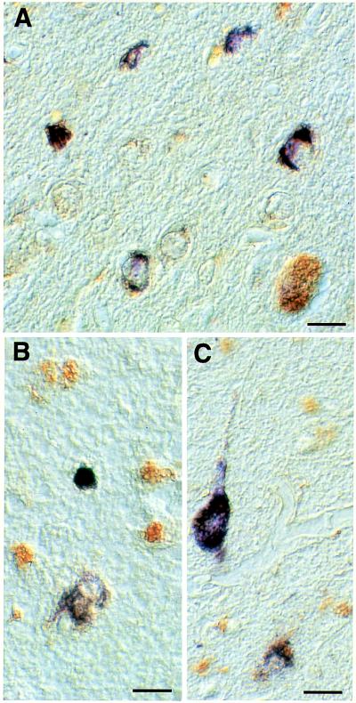
Figure 2.
Temporal cortex from a familial MSTD patient double-stained with the phosphorylation-dependent anti-tau antibody AT8 and the anti-heparan sulfate antibody 10E4. AT8 staining is shown in blue, and 10E4 staining is shown in brown (Nomarski optics). Note staining of some nerve cells and glial cells with 10E4 and double-labeling of nerve cells and glial cells with both AT8 and 10E4. (Bar = 23.5 μm in A, 21 μm in B, and 20 μm in C.)

