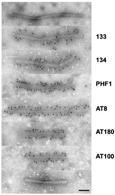
Figure 3.
Immunoelectron microscopy of isolated filaments from familial MSTD brain. The top and bottom panels are unlabeled, and the others are immunogold decorated with phosphorylation-dependent (AT100, AT180, AT8, and PHF1) and phosphorylation-independent (BR133 and BR134) anti-tau antibodies. (Bar = 100 nm.)

