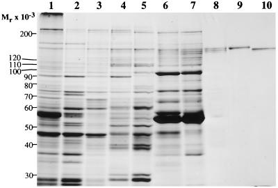
Figure 1.
SDS/PAGE of fractions obtained during the purification of IPF. The starting material and fractions containing the peak inhibitory activity from the various purification steps were dissociated by boiling for 2 min in the presence of 1% SDS/5% 2-mercaptoethanol/10% glycerol/63 mM Tris·HCl, pH 6.8 and were subjected to electrophoresis on an SDS/7.5% polyacrylamide gel. Gel staining was with Coomassie brilliant blue. Lanes: 1, 40 μg of calf-brain homogenate; 2, 40 μg of crude cytosol; 3, 40 μg of 45% ammonium sulfate precipitate; 4, 40 μg of PAE 0.5 M NaCl eluate; 5, 40 μg of HAP 0.05 M phosphate eluate; 6, 40 μg of Yellow-86 0.3 M NaCl eluate; 7, 20 μg of gel filtration peak; 8, 5 μg of sucrose gradient peak; 9, 1.5 μg of Mono Q-purified IPF α; 10, 1.5 μg of Mono Q-purified IPF β.

