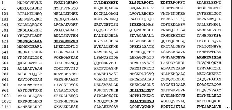
Figure 4.
Comparison of partial IPF α sequences with the sequence of human α fodrin. Amino acid sequences determined for IPF α are shown in boldface type within the initial 1,200 aa residues of human α fodrin as determined by Moon and McMahon (35). The 20-mer beginning with Tyr-26 represents the N terminus of IPF α. The four internal sequences were determined by sequencing peptides produced by proteolytic digestion of IPF α. The highlighted bond between Tyr-1,176 and Gly-1,177 represents the cleavage site for calpain (36).

