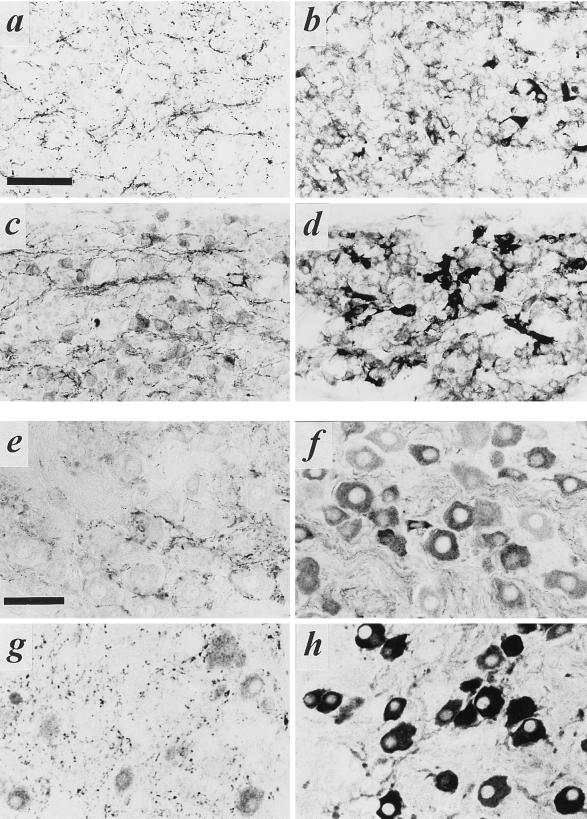
Figure 2.
VAChT and VMAT2 expression in embryonic and adult superior cervical and stellate sympathetic ganglia. (a–d) Staining of superior cervical (a and b) and stellate (c and d) ganglia for VAChT (a and c) or VMAT2 (b and d) on E16. Note copious cholinergic preganglionic terminal immunoreactivity in both ganglia at this stage of development, large numbers of VMAT2-positive principal ganglion cells in both ganglia, and frequent VAChT-positive principal ganglion cells in stellate (c) but not superior cervical (a) ganglion at this time. (e–h) Staining of superior cervical (e and f) and stellate (g and h) ganglia for VAChT (e and g) or VMAT2 (f and h) in adult. Note copious cholinergic preganglionic terminal immunoreactivity in both ganglia, large numbers of VMAT2-positive principal ganglion cells in both ganglia, and frequent VAChT-positive principal ganglion cells in stellate (g) but not superior cervical (e) ganglion at this time. (Bars in a and e = 50 μm; all panels at same magnification.)

