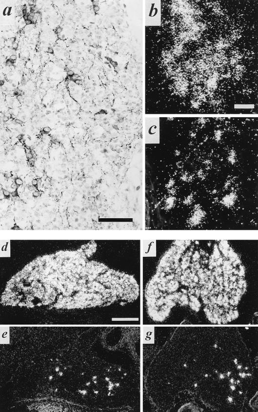
Figure 3.
VAChT/ChAT expression in a subpopulation of principal cells of the developing stellate ganglion. (a) Immunohistochemistry for VAChT in stellate ganglion at E19. Note numerous VAChT-positive principal cells as in Fig. 2, as well as VAChT-positive preganglionic terminals at this stage of development. (b and c) Detection of VAChT mRNA (b) and ChAT mRNA (c) in E19 stellate ganglion. Exposure for 14 days following in situ hybridization. Note b and c represent nonadjacent sections from the same ganglion. (d and e) VMAT2 (d) and VAChT (e) in situ hybridization histochemistry, postnatal day 2 (P2). (f and g) VMAT2 (f) and VAChT (g) in situ hybridization histochemistry, P11. Note relative constancy in the number of VAChT mRNA-positive cells per section of whole ganglion throughout development at E19 (b), P2 (e), and P11 (g). [Bars = 50 μm (a), 10 μm (b and c), and 250 μm (d–g).]

