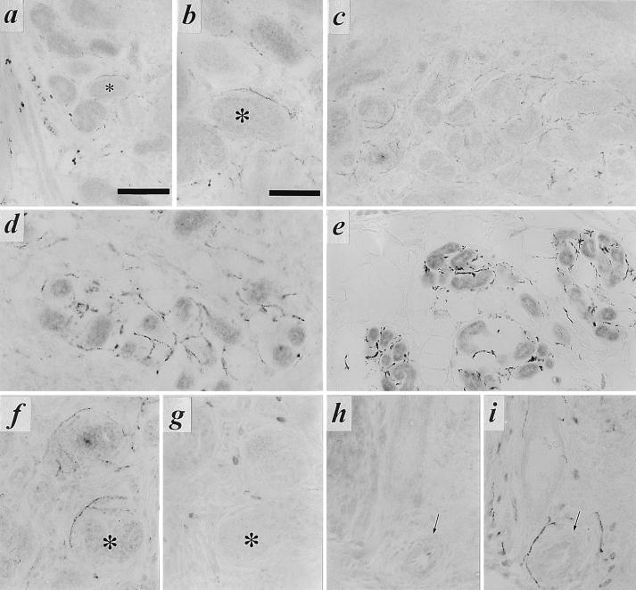
Figure 4.
Expression of VAChT in nerve terminals innervating developing sweat glands in early postnatal development, and mutual exclusivity of VMAT2 and VAChT immunoreactivity in footpad nerve terminals. (a) Immunohistochemical staining for VAChT (as described in legend to Fig. 1) in rat sweat gland tissue at P4. Note intense VAChT-positive neuromuscular junctions within skeletal muscle at bottom left. Faint but distinct terminal staining can be seen on most of the sweat gland epithelial cell clusters, beginning to coil into sweat glands proper at P4. Nonspecific staining of blood cells is seen within lumen of large vessel separating skeletal muscle and sweat gland epithelial regions. Asterisk marks sweat gland seen at high power in b. (b) VAChT-positive terminals at P4, high magnification. Note VAChT-immunoreactivity in terminals closely contacting developing secretory coils of developing eccrine sweat glands. (c–e) VAChT-positive terminals in P8 (c), P11 (d), and adult (e) forepaw sweat glands. Note increasing density of cholinergic terminals through the first two postnatal weeks. (f–i) VAChT (f and h) and VMAT2 (g and i) in adjacent sections (f and g; h and i) of P8 sweat gland. Asterisk in f and g marks sweat gland positive for VAChT (f) and negative for VMAT2 in adjacent section (g). Arrows in h and i mark blood vessel negative for VAChT (h) and positive for VMAT2 (i). VAChT immunoreactivity in sweat gland nerve terminals was shown to be specific by complete loss of immunoreactivity upon preincubation of anti-VAChT antibody, at its final working dilution, with the C-terminal VAChT peptide against which the antibody was raised, at a final concentration of 10 μM. VMAT2 immunostaining was unaffected by preincubation with this concentration of the VAChT peptide. [Bars = 100 μm (a, c, and e) and 50 μm (b, d, and f–i).]

