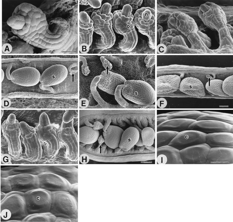
Figure 2.
Cryo-scanning electron microscopy micrographs of ovules and seeds of fis mutants and fertilized wild-type plants. Developing ovules [nucellar column (n) protruding from the inner integument (ii) and the outer integument (oi) as shown in B] of (A) wild-type, (B) fis1/fis1 homozygote, (C) fis2/fis2 homozygote, and (G) FIS3/fis3 heterozygote. (D) Sexually fertilized seeds(s) of pi/pi FIS/FIS plants 7 days after fertilization. Unfertilized ovules shrivel (arrow). Seeds developing without fertilization(s) of (E) fis1/fis1 homozygote, (F) fis2/fis2 homozygote, and (H) FIS3/fis3 heterozygote. Collumella (c) on the surface of (I) sexually fertilized seed of wild type and (J) autonomously developing fis2 homozygous seeds. (Bar = 20 μm for A–C, G, I, and J, 100 μm for D–F, and 200 μm for H.)

