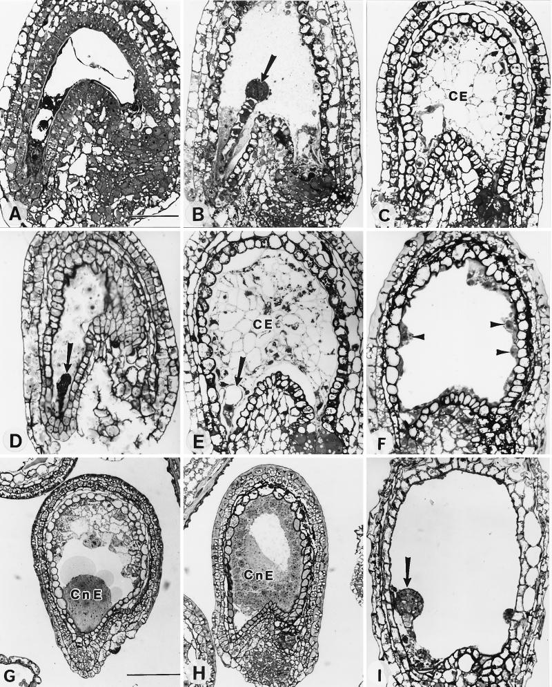
Figure 3.
Endosperm and embryo development of wild-type and fis seeds. (A) Longitudinal section through an ovule of an unfertilized pistillata (pi/pi) mutant. No endosperm or embryo development is visible. (B) Longitudinal section through an ovule of a 0.6-mm fruit from a pi/pi homozygote 3 days after fertilization with FIS/FIS pollen showing embryo (arrow). (C and D) Sections through an autonomously developing fis1 seed from a FIS1/fis1 heterozygous plant (from a 0.8-mm fruit). Notice cellularized endosperm (CE) and the embryo-like structure (D, arrow). (E) Section through an autonomously developing fis2 seed from a FIS2/fis2 heterozygote showing cellularized endosperm (CE) and a zygote-like structure (arrow) (from a 0.8-mm fruit). (F) Section through an autonomously developing fis3 seed from a 0.9-mm fruit from a FIS3/fis3 heterozygote showing free nuclear endosperm (arrowheads). (G and H) Autonomous endosperm development in a fis1/fis1 homozygote showing free nuclear coenocytic endosperm (CnE) (from a 1-mm fruit). (I) Embryo arrested at the globular stage 5 days after pollination of a fis1/fis1 homozygous plant with FIS/FIS pollen (arrow) (from a 1.2-mm fruit). (Bar = 50 μm for A–F and I and 100 μm for G and H.)

