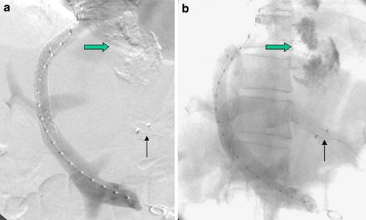
Fig. 2.

The same patient as in Fig. 1 immediately following TIPS creation and AVP embolization of the coronary vein (a; thin arrow). The unsubtracted image (b) shows the radiopaque sclerosing agent from prior endoscopic sclerotherapy (fat arrow) in the esophageal varices
