
Figure 2.
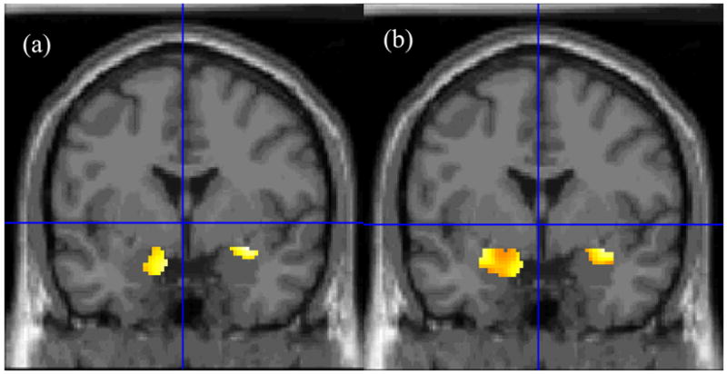
Statistical maps of voxel-wise between-group comparisons of amygdala activation during the fear rating condition relative to the passive viewing condition during the presentation of (a) fearful and (b) happy faces. BI adolescents compared to BN adolescents exhibited greater bilateral amygdala activation to both fearful and happy faces. [Coordinates for fearful faces: 32 –2 –12; happy faces: −28 –4 –12].