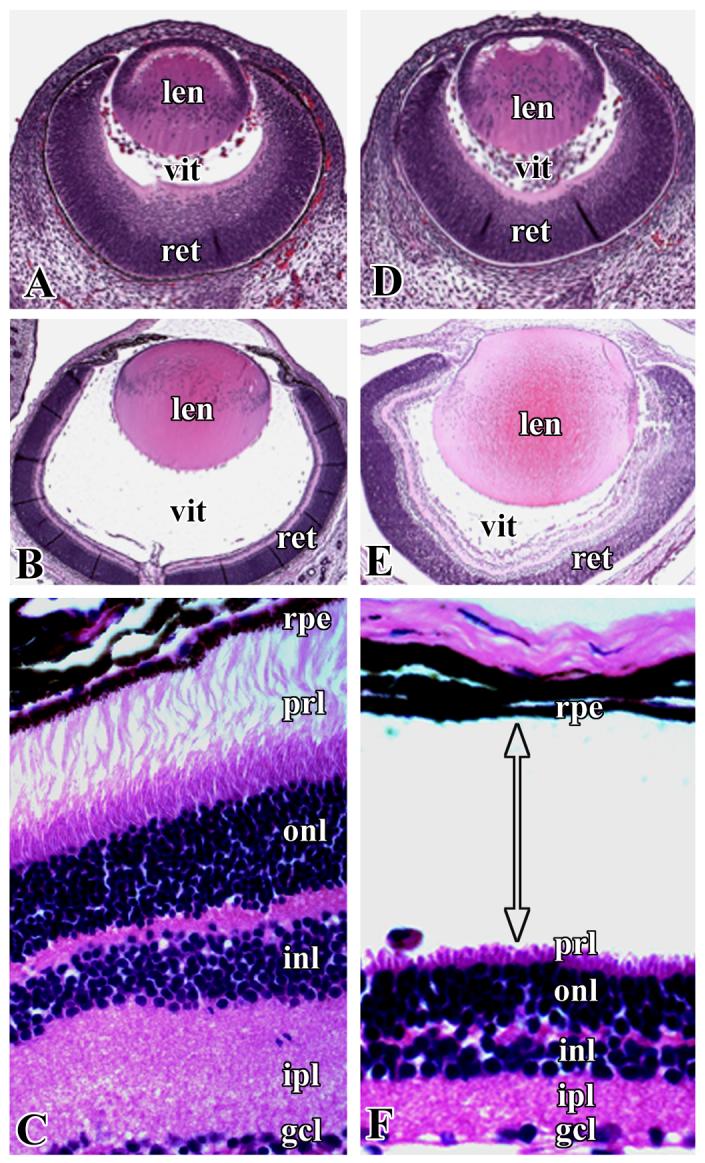
Figure 1.

Representative pictures of H&E stained sections of the eyes of wild-type (A, B, C) and cKO (D, E, F) mice. A and D, embryonic age 14.5 days; B and E, postnatal day 2; C and F, retinal sections of wild-type and cKO mice, respectively, shown at higher magnification to illustrate retinal detachment at the pigment epithelia and photoreceptor layer. len- lens, vit- vitreous, ret- retina, rpe- retinal pigment epithelium, prl- photoreceptor layer, onl- outer nuclear layer, inl- inner nuclear layer, ipl- inner plexiform layer, gcl- ganglion cell layer. Arrow with double heads indicates the position of retinal detachment.
