Figure 3.
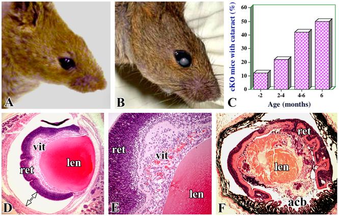
Cataract development in cKO mice. Representative pictures of wild-type (A) and cKO (B) mice show the macroscopic cataracts. (C) The incidence of macroscopic development of cataracts in one or both eyes in cKO mice in relation to age. Representative photographs of eye sections of cKO mice illustrate retinal detachment (D) presence of inflammatory and blood cells in the vitreous (E), and the disorganized appearance of a cataractous lens (F). acb- anterior chamber, other labels are similar to those indicated in Figure 2.

