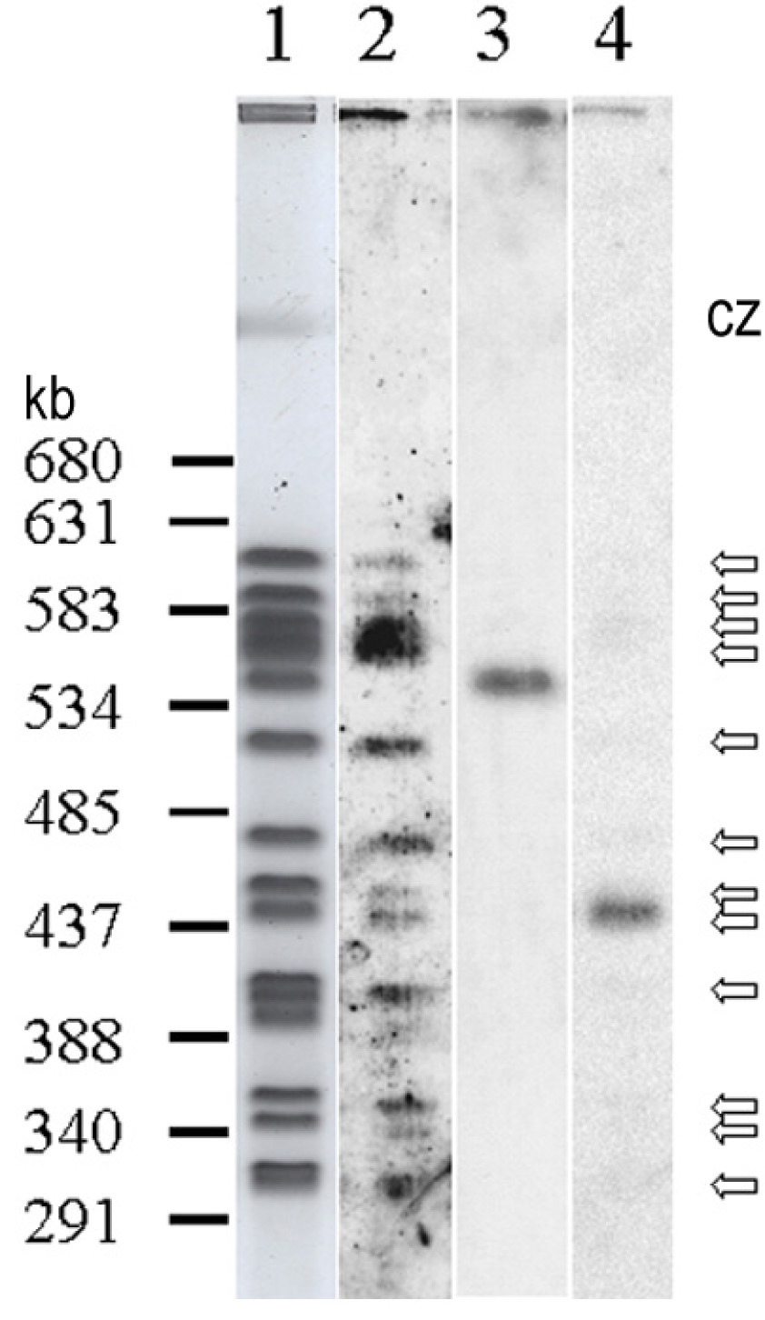
Figure 4. Hybridization mapped the sequence encoding UCS to a single P. murina chromosome.

Lane 1: Negative image of Sybr-gold stained chromosomes resolved by CHEF. Lanes 2–4, autoradiograms showing chromosomes that hybridized to probes for MSG, kex1 and UCS, respectively. Numbers to the left of lane 1 indicate locations of markers ranging from 291 to 680 kilobasepairs (markers not shown). Open arrows to the right of lane 4 indicate chromosome bands scored as positive for the MSG probe. CZ indicates the compression zone, where DNA from the mouse host lung is located (visible in lane 1).
