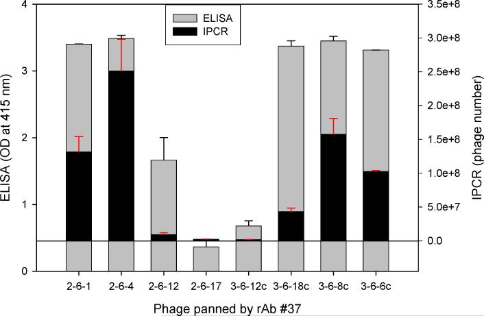
Fig. 3.
Comparison of (RT) IPCR and traditional ELISA in determining phage binding to a single MS rAb. Eight different phage (5 × 10⁸) were added to ELISA wells pre-coated with 50 ng of MS rAb #37 and binding was assayed by RT-IPCR or standard ELISA using anti-M13 antibody as described in the Fig. 2 legend. White and black bars show ELISA OD values and IPCR bound phage number, respectively, for the same phage population. With 5 × 10⁸ phage, ELISA values for phage 2−6−1, 2−6−4, 3−6−18c, 3−6−8c and 3−6−6c were similar, in contrast to distinct differences in the number of bound phage revealed by IPCR. Error bars represent standard deviation of duplicate samples.

