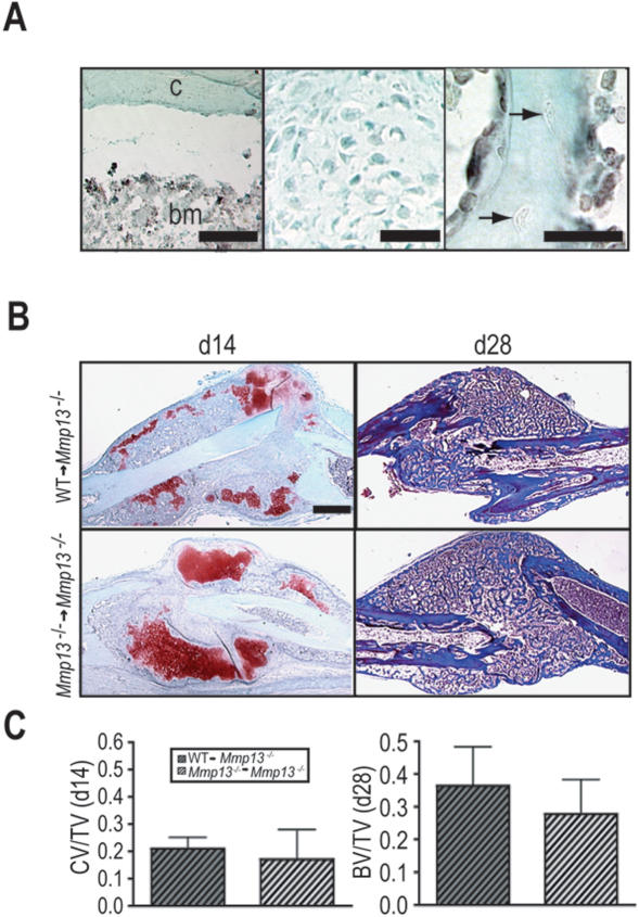
Figure 5. Transplant of WT bone marrow does not rescue the Mmp13 −/− non-stabilized fracture healing phenotype.
(A) Immunostaining for GFP on callus tissues from Mmp13−/− mice transplanted with bone marrow from β-actin GFP mice (GFP→Mmp13−/− mice) . (Left panel) Bone marrow cells (bm) are positive for GFP (black staining) showing they are donor-derived but the adjacent cortex (c) is negative. (Middle panel) Chondrocytes at day 14 and (Right panel) osteocytes embedded in the new bone (arrows) at day 28 do not stain for GFP, showing they are host-derived. (B, Left column) SO and (Right column) Masson's Trichrome staining of non-stabilized fracture calluses from Mmp13 −/− mice transplanted with WT bone marrow (WT → Mmp13 −/−) and Mmp13 −/− mice transplanted with Mmp13 −/− bone marrow (Mmp13 −/− → Mmp13 −/−) show no difference in the amount of cartilage volume at 14 days post-fracture (WT → Mmp13−/− n = 6, Mmp13−/−→ Mmp13−/− n = 5) and no difference in the amount bone at day 28 (WT → Mmp13−/− n = 7, Mmp13−/−→ Mmp13−/− n = 4). (C) Histomorphometric analyses of total cartilage volume as a proportion of total callus volume (CV/TV; day 14) and total bone volume as a proportion of total callus volume (BV/TV; day 28) demonstrate no significant difference between WT → Mmp13 −/− and Mmp13 −/− → Mmp13 −/− animals, suggesting that bone marrow transplant does not rescue the Mmp13 −/ non-stabilized fracture healing phenotype. Bonferroni corrected t-test, bars represent means ± SD. Scale bars: (A, left and middle) = 50 µm, (A, right) = 25 µm, B = 1 mm.

