Figure 2.
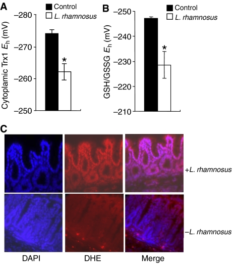
In vivo changes in intracellular redox in mouse intestinal epithelial cells contacted by commensal bacteria. (A) Lactobacillus-induced Trx1 oxidation in epithelial cells as analyzed by redox Western analysis. The graph shows the Eh values for Trx1 in proximal jejunum 30 min after treatment with a bolus of bacteria (open bars) as compared with untreated controls (filled bars). Data are represented as means±s.e. Asterisks denote a statistically significant difference from control (P<0.05). (B) Lactobacillus-induced changes in the redox pools of GSH/GSSG. The graph shows the Eh values for GSH/GSSG in the jejunum 30 min after treatment with a bolus of bacteria (open bars) as compared with untreated controls (filled bars). Data are represented as means±s.e. The asterisks denote a statistically significant difference from control (P<0.05). (C) In situ detection of superoxide in mouse colon treated with Lactobacillus. After 30 min of infusion of Lactobacillus, mouse colon was stained with DHE and imaged as described. Under identical tissue processing and imaging conditions, nuclear fluorescence in mouse colon treated with bacteria (top panels) is markedly increased compared with colon from untreated control mouse (bottom panels).

