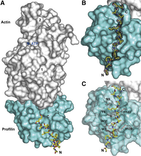
Figure 3.
Crystal structure at 1.8-Å resolution of the ternary complex of profilin–actin with the loading poly-Pro site of human VASP. (A) General view of the structure (actin, gray; profilin, cyan; VASP peptide, all-atom representation). The view is rotated ∼90° relative to the ‘classical' orientation of actin shown in Supplementary Figure 3A. (B) Electron density map around the VASP peptide, contoured at 1.0σ. (C) Illustration of the main interactions of the VASP peptide with profilin amino acids (colored red, under a transparent surface representation of profilin).

