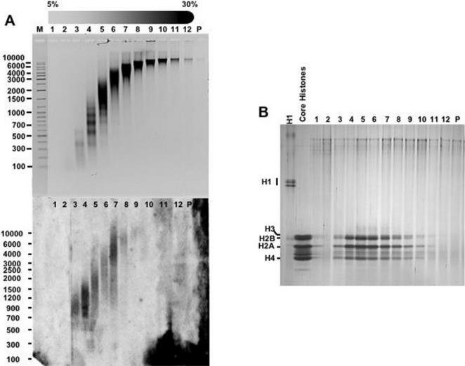
Figure 2. RNA remains associated to chromatin at high ionic strength.
A) Chicken liver chromatin was prepared by micrococcal nuclease digestion of purified nuclei and then subjected to sedimentation through a linear 5%–30% sucrose gradients containing 0,65 M NaCl. After centrifugation, fractions were collected and their DNA (top) and RNA (bottom) content determined as in Figure 1. Fraction numbers are indicated. Lane M, corresponds to molecular weight markers. B) The histone content of each fraction was analyzed by SDS-PAGE (lanes 1-P). As controls, H1 from calf thymus and hydroxylapatite-purified core histones from chicken liver are also presented. The gel was stained with silver.

