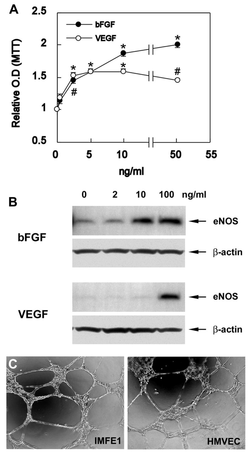
Fig 3. Responsiveness of IMFE1 cells to angiogenic growth factors and growth on Matrigel.

(A) Growth response curves of IMFE1 cells 72hs after stimulation with bFGF and VEGF. Results are expressed as a ratio of the absorbance of the stimulated wells to the absorbance of the non-stimulated wells. Data are presented as mean ± SE (n = 6). (*P<0.0003, #P<0.002, stimulated wells compared with non-stimulated wells, two-tailed Student’s t test) (B) eNOS expression analyzed by Western blot. IMFE1 cells were incubated with medium containing 0–100 ng/ml VEGF or bFGF for 48 hr. β-Actin was used as loading control. (C) Tube formation assay. IMFE1 cells grown on Matrigel extracellular matrix formed tube-like structures that were similar to those formed by primary HMVEC.
