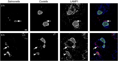
Figure 6: SCVs fuse with preformed C. burnetii parasitophorous vacuoles in co-infected cells.
HeLa cells were infected with C. burnetii for 48 h before infection with wt Salmonella. At 2 or 6 h p.i., cells were fixed and processed for immunofluorescence microscopy using α-Salmonella LPS (red), α-Coxiella (green) and α-LAMP1 (blue) antibodies. Projections of 12–15 confocal planes are shown. Arrows indicate Salmonella that are adjacent to, but not within, a Coxiella vacuole. An arrowhead indicates a vacuole that contains both Salmonella and Coxiella. Scale bar = 5 μm.

