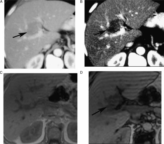
Figure 1. .
Contrast-enhanced (CT, portal phase) (A, B), in-phase (C), opposed phase (D), showing indeterminate CT focal liver lesion (arrow) that is well characterized by MRI as focal fatty infiltration (drop-off signal on opposed phase chemical shift relative to in-phase), in a 63-year-old female complaining of breast cancer, who underwent CT for assessment of presence of metastases.

