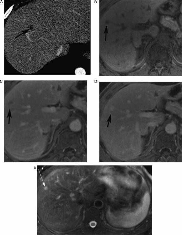
Figure 2. .
Contrast-enhanced (CT, portal phase) (A), pre-contrast VIBE (B), arterial phase VIBE (C), portal phase (D), and T2 IR (E), showing indeterminate CT focal liver lesion that is well characterized by MRI as hemangioma (peripheral puddling on early contrast phase, filling up on later phase and very bright signal on T2), in a 43-year-old male.

