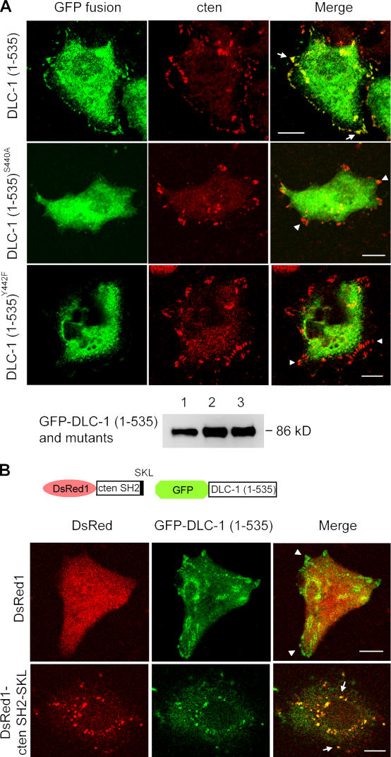
Figure 3.
Subcellular localization of DLC-1 and its recruitment by cten SH2. (A) A549 cells grown on coverslips were transfected with pEGFP–DLC-1 (1–535), pEGFP–DLC-1 (1–535)S440A, or pEGFP–DLC-1 (1–535)Y442F. After labeling with anti-cten antibodies followed by Alexa Fluor 594–conjugated secondary antibody, cells were visualized with a confocal microscope. Arrows indicate cten and GFP fusion protein colocalized at focal adhesions. Arrowheads show only cten at focal adhesions. About 100 GFP-positive cells were examined in each transfection. More than 90% of GFP-positive cells showed focal adhesion localization when transfected with GFP–DLC-1 (1–535), and no GFP-positive cells transfected with DLC-1 mutants showed focal adhesion localization. Cell lysates from A549 transiently expressing GFP–DLC-1 (1–535; lane 1), GFP–DLC-1(1–535)S440A (lane 2), or GFP–DLC-1 (1–535)Y442F (lane 3) were immunoprecipitated with anti-GFP and analyzed by immunoblotting with anti-GFP to show similar amounts, and correct sizes of recombinant proteins were expressed. (B) A549 cells grown on coverslips were cotransfected with pDsRed1/pEGFP–DLC-1 (1–535) or pDsRed1-cten SH2-SKL/pEGFP-DLC-1 (1–535). Cells were visualized with a confocal microscope. Note that the DsRed-cten SH2-SKL and GFP–DLC-1 (1–535) were colocalized at peroxisomes (arrows), although some GFP–DLC-1 (1–535) proteins were still detected at focal adhesions (at different focus plane; not depicted), as predicted, because of the presence of endogenous tensins in A549 cells. Arrowheads show only GFP–DLC-1 (1–535) at focal adhesions. Bars, 10 μm.

