Figure 2.
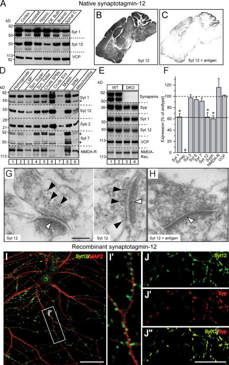
Expression and localization of synaptotagmin-12. (A) Total homogenates of cortex, cerebellum, hippocampus, olfactory bulb, brain stem, and spinal cord isolated from the brains of adult rats were analyzed with antibodies to synaptotagmin-12, -1, and VCP (loading control). 20 μg of total protein was loaded into each lane. Note that synaptotagmin-12 is expressed at different levels in various brain regions, with the highest expression level in cerebellum. (B and C) Sections of brains isolated from adult mice were labeled with antibody to synaptotagmin-12. (B) Labeling of brain sections indicates that synaptotagmin-12 is abundantly expressed in various brain regions. (C) Brain sections were labeled with antibody to synaptotagmin-12 mixed with ∼1 mg/ml recombinant GST-linker fusion protein that was used for immunization. Note that synaptotagmin-12 immunoreactivity is greatly reduced in the presence of the antigen. (D) Distribution of native synaptotagmin-12 in subcellular fractions of adult brain separated by centrifugation. Similar to synaptotagmin-1 (Syt 1) and synaptobrevin/VAMP2 (Syb 2), synaptotagmin-12 is enriched in synaptic vesicle fraction (crude SVs). Asterisks indicate proteolitically cleaved fragment of synaptotagmin-1 and various splice variants of synaptotagmin-7. (E) Expression of indicated proteins in brains of adult wild-type and synapsin double-knockout mice (DKO) was analyzed by quantitative immunoblotting with I125-labeled secondary antibodies. (F) The relative expression levels of indicated proteins in DKO brains were normalized to VCP and presented as the percentage of wild type. Note that expression of synaptotagmin-12 and other synaptic vesicle proteins is reduced by ∼40%. Data are shown as the mean ± the SEM. (G and H) Electron micrographs of adult mouse hippocampal sections labeled with synaptotagmin-12 antibody alone (G) or in the presence of ∼1 mg/ml of the antigen (synaptotagmin-12 linker region) used for immunization (H). The postsynaptic densities are marked by white arrowheads. Note that synaptotagmin-12 immunoreactivity is restricted to presynaptic sites (G; black arrowheads) and inhibited in the presence of the antigen (H). (I and J) Localization of recombinant synaptotagmin-12 in cultured hippocampal neurons. The neurons were infected at 5 d after plating (5 DIV) with lentivirus expressing full-length synaptotagmin-12 cDNA, and double labeled at 14 DIV with antibodies to synaptotagmin-12 and the dendritic marker MAP2 (I; Syt 12, green; MAP2, red) or Syt 12 and synaptic marker synaptophysin (J; Syt 12, green; synaptophysin, red). Inset (I′) illustrates enlarged fragment of dendrite marked by white box. Note that recombinant synaptotagmin-12 is concentrated in discrete clusters apposed to dendrites and colocalized with synaptophysin. Bars: (G) 100 nM; (I and J") 20 μM.

