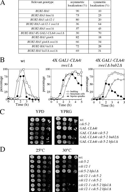
Figure 6.
Disappearance of Bub2 from the mother-bound SPB depends on proteins localized at the bud neck. (A) Localization of Bub2-HA3 was analyzed on formaldehyde-fixed cells undergoing properly oriented anaphase (as assessed by DAPI staining) from logarithmically growing cultures at 25°C, with the exception of BUB2-HA3 4× GAL1-CLA4t swe1Δ cells, which were grown in raffinose-containing medium, arrested in G1 by α factor, and released in the presence of galactose for 3 h to induce GAL1-CLA4t expression. At least 150 anaphase cells were scored for each strain. (B) Wild-type (ySP41), 4× GAL1-CLA4t swe1Δ (ySP2711), and 4× GAL1-CLA4t swe1Δ bub2Δ (ySP2728) cells were grown in YEPR at 25°C, arrested in G1 with α factor, and released in YEPRG at 25°C. Galactose was added 30 min before the release. 120 min after the release, 10 μg/ml α factor was readded to prevent cells from entering a second cell cycle. Cells were collected at the indicated times for kinetics of budding, nuclear division, and bipolar spindle formation/breakdown after in situ immunostaining of tubulin. Bipolar spindles include both metaphase and anaphase spindles. (C) Serial dilutions of wild-type (ySP41), cdc5-2 (ySP324), GAL1-CLA4t (ySP2622), GAL1-CLA4t cdc5-2 (ySP3565), GAL1-CLA4t cdc5-2 bub2Δ (ySP4802), and GAL1-CLA4t cdc5-2 bfa1Δ (ySP4804) cell cultures were spotted on either YPD (GAL1 promoter off) or YPRG (GAL1 promoter on) plates and incubated at 25°C for 3 d. (D) Serial dilutions of wild type (ySP41), cdc5-2 (ySP324), cdc12-1 (ySP293), cdc5-2 bfa1Δ (ySP3671), cdc12-1 cdc5-2 (ySP4473 and ySP4474), and cdc12-1 cdc5-2 bfa1Δ (ySP4518 and ySP4519) were spotted on YPD plates, which were incubated at 25 and 30°C for 2 d.

