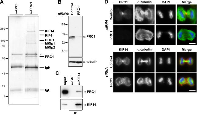
Figure 1.
The central spindle MAP PRC1 interacts with multiple kinesins. (A) Proteins immune precipitated from 10 mg of anaphase HeLa cell extract with affinity-purified rabbit antibodies to GST or PRC1 were separated on minigels and identified by mass spectrometry. The immunoglobulin G heavy (IgH) and light chains (IgL) are marked, and the asterisk denotes a nonspecific contaminant found with both control and KIF14 antibodies. (B) Lysates from HeLa cells treated with control or PRC1 siRNA duplexes for 36 h were Western blotted for PRC1 and α-tubulin as a loading control. (C) GST and KIF14 immune precipitates were Western blotted for KIF14 and PRC1. (D) HeLa cells treated with control or PRC1 siRNA duplexes for 30 h were fixed and stained with antibodies to PRC1 or KIF14 (red) and α-tubulin (green). DNA was stained with DAPI (blue). Bars, 10 μm.

