Figure 3.
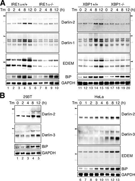
Involvement of the IRE1–XBP1 pathway in the induction of Derlin-1 and -2 in response to ER stress. (A) IRE1α+/+, IRE1α−/−, XBP1+/+, and XBP1−/− MEFs were treated with 10 μg/ml tunicamycin (Tm) for the indicated periods. Total RNAs were isolated and analyzed by Northern blot hybridization using a DIG-labeled cDNA probe specific to mouse Derlin-2, Derlin-1, EDEM, BiP, or GAPDH. Closed and open arrowheads indicate the migration positions of 28S ribosomal RNA (4.7 kb) and 18S ribosomal RNA (1.9 kb), respectively. (B) 293T or HeLa cells were treated with 2 μg/ml tunicamycin (Tm) for the indicated periods. Total RNAs were analyzed as in A using a DIG-labeled cDNA probe specific to human Derlin-2, Derlin-3, EDEM, BiP, or GAPDH.

