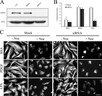
Figure 9.
mDia1 cooperates with PAK1 in LPA regulation of collagen matrix contraction silencing in human fibroblasts. (A) Cells were transfected for 12 h with 700 nM siRNA or sense RNA only (Mock) and cultured an additional 24 h in growth medium without siRNA. Extracts were prepared and subjected to SDS-PAGE and immunoblotted to analyze levels of mDia1 and actin. (B) mDia1-silenced and mock-transfected cells were harvested and used to prepare floating collagen matrices. Samples were incubated for 4 h in DME with 5 mg/ml BSA and 50 ng/ml PDGF or 10 μM LPA added as shown. At the end of the incubations, samples were fixed, and the extent of matrix contraction was measured as the decrease in matrix diameter. Data shown are arithmetic means ± SD for three separate experiments. (C) At the end of the transfection period, mock- and siRNA-transfected cells were incubated in serum-free medium for 36 h, followed by 4 h in DME with 5 mg/ml BSA and 50 ng/ml PDGF or 10 μM LPA added as shown. Stable microtubules were detected as previously described (Gundersen et al., 1994; Cook et al., 1998). Subsequently, the indicated samples were treated with 2 μM nocodazole for 2 h . After two rinses with microtubule-stabilizing buffer (MSB; 85 mM Pipes, pH 6.9, 1 mM EGTA, 1 mM MgCl2, 2 M glycerol, 1 μg/ml leupeptin, 1 μg/ml pepstatin A, and 1 mM 4-(2-aminoethyl)-benzenesulfonyl fluoride), samples were treated with 1 ml of MSB containing 200 μg/ml saponin for 5 min at 37°C to extract tubulin monomer, rinsed with MSB, and fixed with methanol (−20°C) for 10 min, and then stained with anti-tubulin antibody. Bar, 50 μm.

