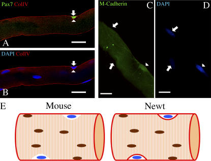
Figure 2.
Satellite cells can be copurified with isolated single skeletal muscle fibers. (A–D) Satellite cells are attached to the myofiber after isolation and plating. (A and B) Arrows point to a Pax7+ satellite cell, and arrowheads point to the collagen IV+ basal lamina. (C and D) Arrows point to two myonuclei, and arrowheads point to an M-cadherin+ satellite cell. (E) Schematic model of mouse and newt myofibers. Satellite cells (blue nuclei) are tightly attached to both mouse and newt myofibers, but an additional basement membrane (maroon) separates the satellite cells from the sarcolemma. Bars, 50 μm.

