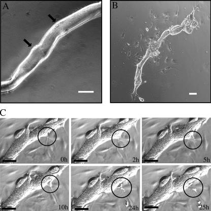
Figure 3.
Progeny cells bud off the myofiber and proliferate. (A) Photomicrograph showing an isolated single newt skeletal muscle fiber directly after attachment. Note the visible striation demarking the sarcomeres. Arrows point to two visible nuclei, which could either be myonuclei or located in satellite cells. (B) Photomicrograph showing the same 15-d-old myofiber in culture. The myofiber morphology has changed and several lobular structures are seen while mononucleate progeny has been produced. (C) Time-lapse photomicrographs showing a sequence of one representative budding event, which leads to the derivation of a mononucleate cell. Note the protrusion of the myofiber in the circled area, which is concomitant with the appearance of a mononucleate progeny. Time points indicate the duration of the one specific budding event. A longer movie capture is shown in Video 1. Video 1 is available at http://ww.jcb.org/cgi/content/full/jcb.200509011. Bars, 50 μm.

