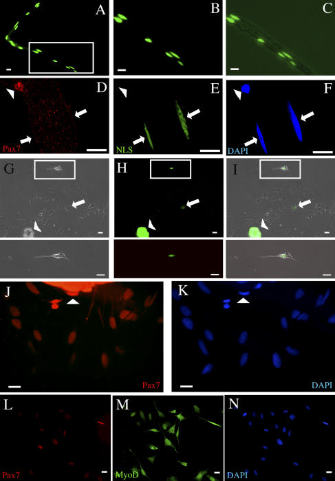
Figure 4.
Myofiber-derived proliferating cells are satellite cell progeny. (A–C) Photomicrographs showing that injected fluorescent NLS-dextran marks the nuclei of the myofiber. B and C are high power magnifications of the boxed area in A. C is an overlay of the fluorescent and light microscopy images. (D–F). Photomicrographs showing that the fluorescent dextran exclusively labels myonuclei in the syncytium, but not the nuclei in satellite cells. Arrows point to mononuclei, arrowheads point to satellite cell. (G–I) Photomicrographs showing that the vast majority of the myofiber progeny lack the NLS-dextran lineage tracer. Although no fluorescent dextran-containing progeny were seen in 69 of 70 single myofiber cultures, these images show the single occasion when two fluorescent dextran+ cells were detected (arrows and boxes), but these cells did not proliferate. Arrowheads point to the myofiber, which has hypercontracted. The pictures underneath G–I are enlarged images of the boxed area. (J and K) Most of the myofiber-derived progeny remain Pax7+ (red) directly after activation, but the intensity of the staining is strongest closest to the hypercontracted myofiber (arrowheads). (L–N) Satellite cell progeny express Pax7 (red) and MyoD (green) for several generations. Bars, 50 μm.

