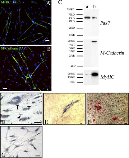
Figure 5.
Newt satellite cell progeny are multipotent. (A and B) Newt satellite cell progeny form myotubes in myogenic media. Note the myosin heavy chain+ myotubes in A and the M-cadherin+ (MyHC) myotubes in B. (C) Western blot analyses show the increased amount of M-cadherin and myosin heavy chain and the reduced amount of Pax7 proteins as a result of myogenic differentiation (lane a, proliferation medium; lane b, after 6 d in myogenic differentiation medium). (D) Satellite cell progeny can enter an adipogenic pathway, as revealed by Oil red staining in lipid droplets (arrowheads). The arrow points to a myotube that is devoid of lipid droplets. The cultures were counterstained by hematoxylin. (E and F) Satellite cell progeny can enter an osteogenic pathway. An alkaline phosphatase+ focus is shown in E, and Alizarin red marks calcium deposits produced by osteogenic cells in F. (G) Lack of Alizarin red staining in cells cultured in proliferation media. Bars, 50 μm.

